
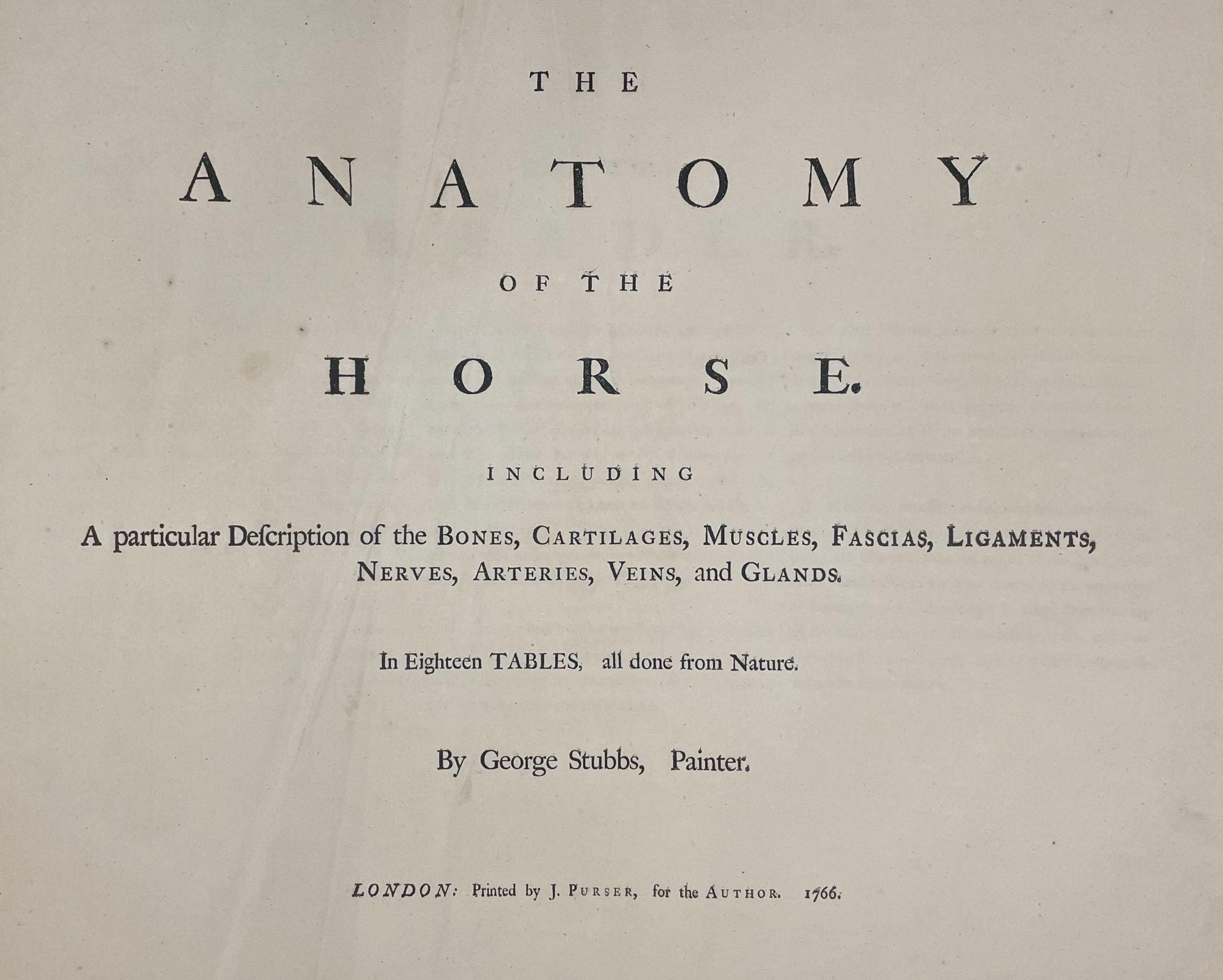
STUBBS, George (1724-1806)
The Anatomy of the Horse: Including a Particular Description of the Bones, Cartilages, Muscles, Fascias. Ligaments, Nerves, Arteries, Veins, and Glands
London: J.Purser for the Author, 1766. Oblong imperial broadsheets (entirely uncut). (19 1/2 x 23 1/4 inches). (2) 47pp. Rare errata leaf pasted onto endpaper. 24 etched plates all by Stubbs.
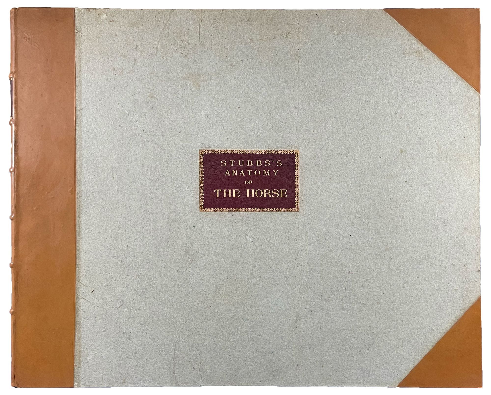
Half calf over gray boards to style, gilt morocco label to upper cover, raised bands, gilt title on label.
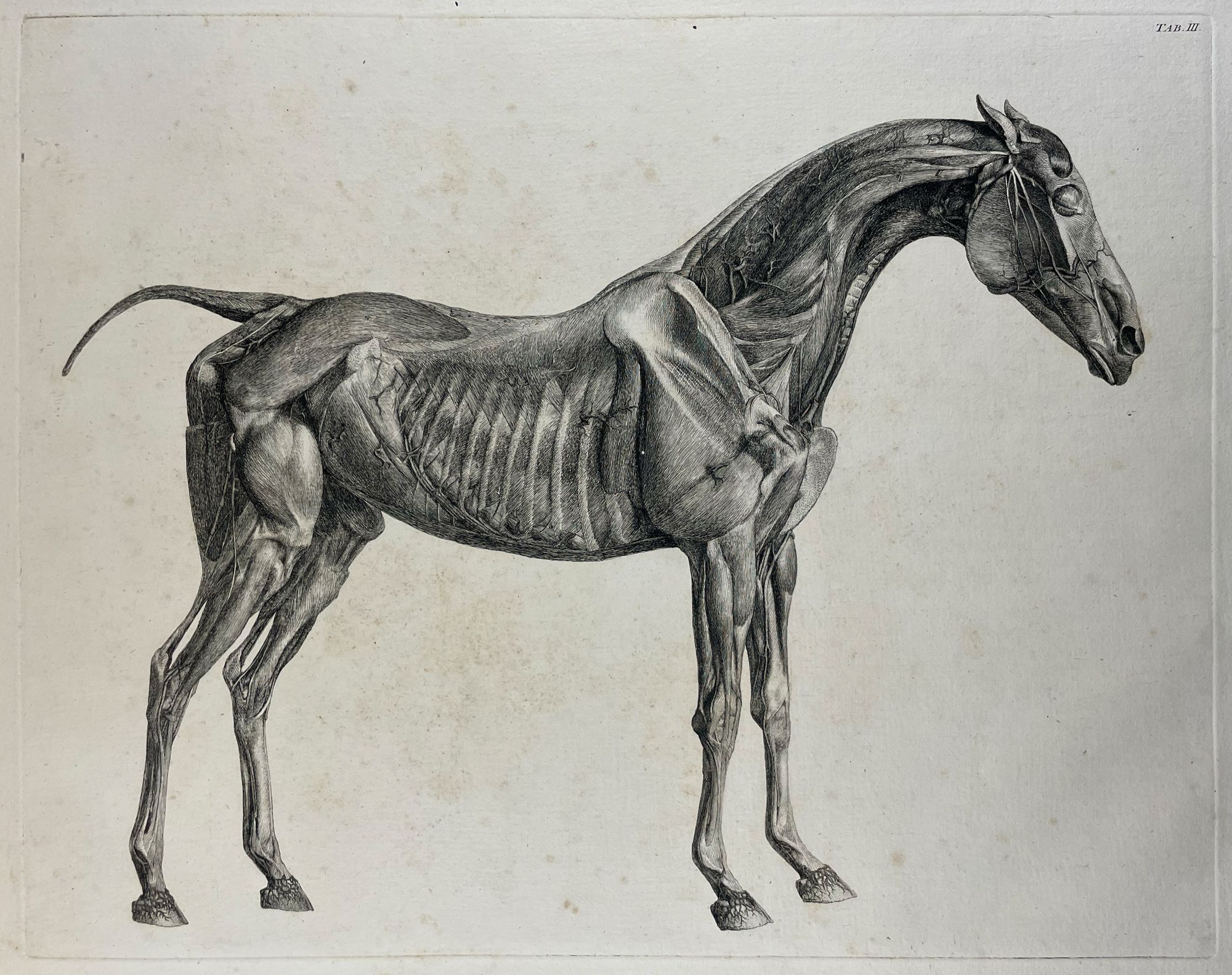
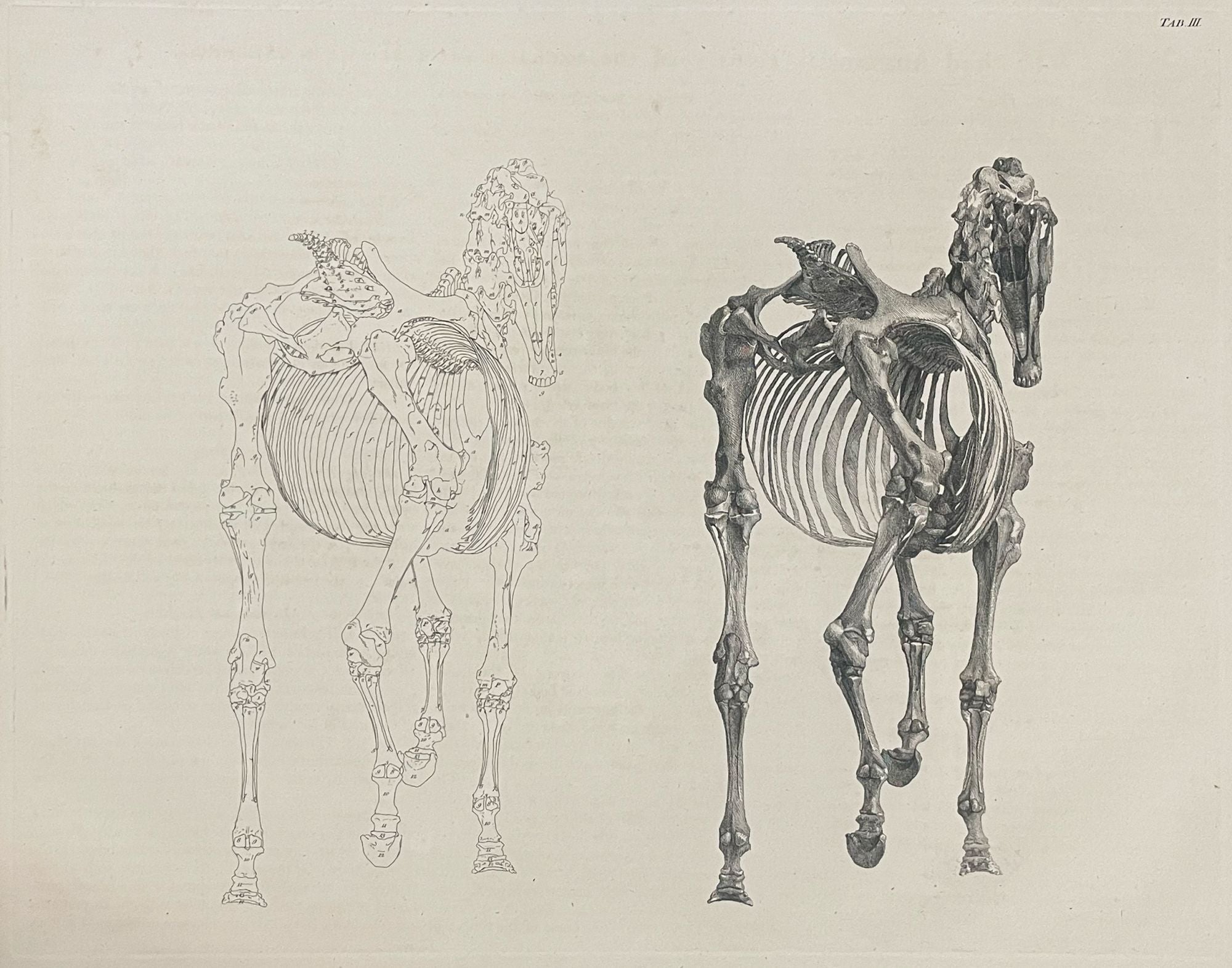
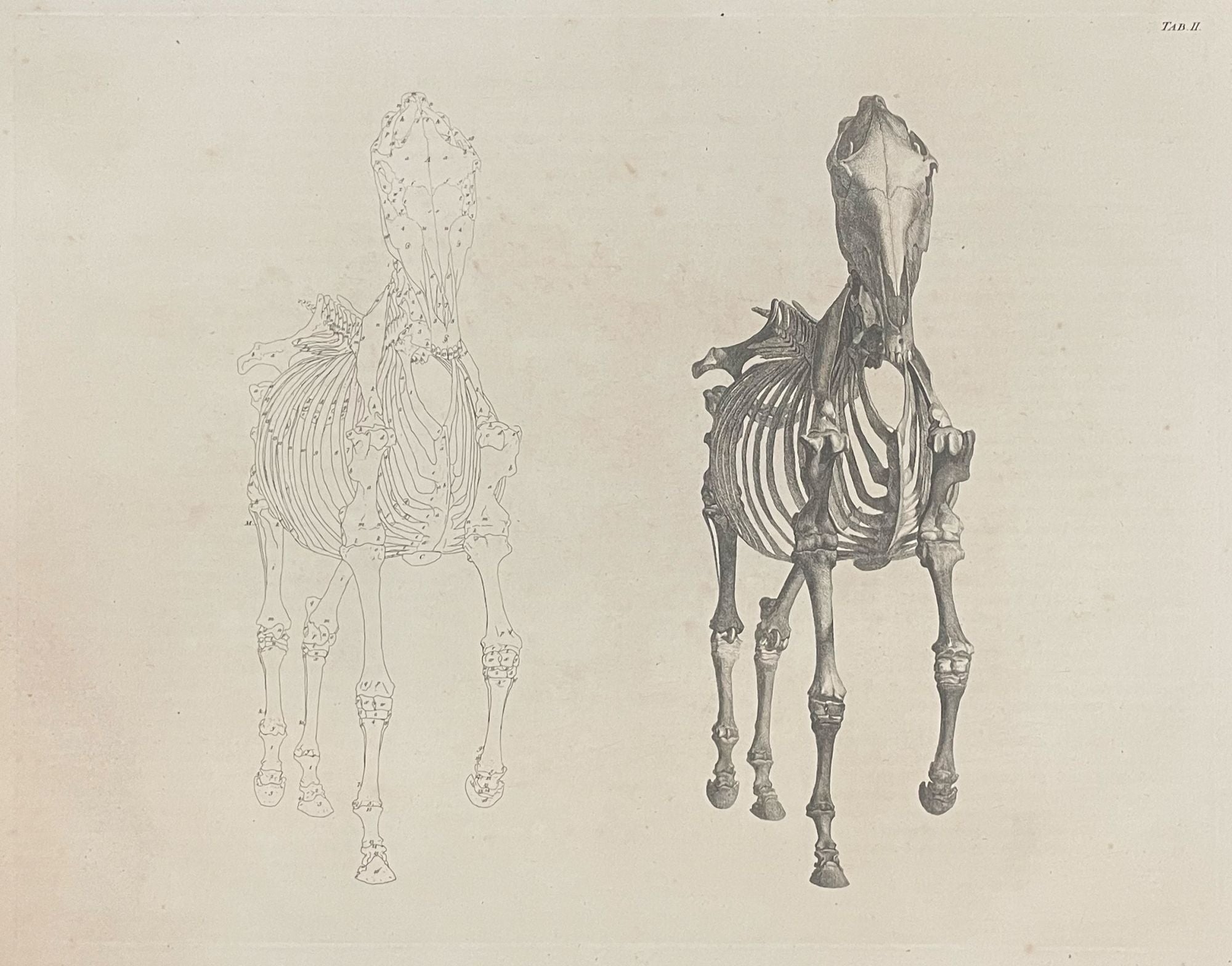
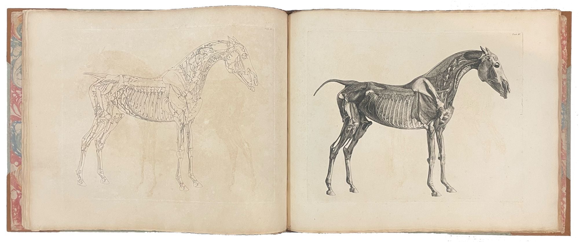
First edition, on laid paper throughout. A landmark work of equine anatomy. One of a select group of books which can be said to have 'revolutionised men's understanding of the natural world' (Lennox-Boyd). The etchings have a 'fine exactness and austere beauty' that 'give them a timeless beauty' (Ray).
The desirability of this work is vastly increased if the plates are on thick laid paper, used for the 1766 first edition. First edition early printings from the plates shows a precision that later impressions, printed on wove paper, cannot match. Examples on laid paper are increasingly difficult to find - even the Duke of Gloucester's copy (sold for over $34,000 at auction in 2006) was printed on wove paper. Stubbs created these remarkable illustrations over a period of 18 months, during which he painstakingly dissected a number of horses, keeping carcasses in his studio for six or seven weeks. Stubbs taught himself how to make the engravings, and produced them over the next six years. The plates document all layers of equine anatomy, revealing the muscles, ligaments, nerves, veins, skeleton, etc. Stubbs' bibliographer Christopher Lennox-Boyd ranked Anatomy of the Horse as 'among the most important of the several works of its time which, by emphasizing the importance of precise systematic observation, revolutionised men's understanding of the natural world.' Originally published in 1766, the work's enduring popularity saw it being issued well into the 19th century.
Garrison and Morton 308.1; Dingley Comben 600: Eales (Cole) 1840; Huth p.42; Lennox-Boyd, Stubbs 165-188; Mellon Books on the Horse and Horsemanship 57; Nissen ZBI 4027; Ray p.6; Sparrow pp.165-188; Brunet, V, p.571; ESTC T147211; Norman 2032 (later issue, plates watermarks '1798').
Item #40544
Price: $30,000.00